
Manual


®
English
Eye – Microscopic Anatomy
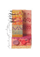
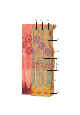
The model illustrates the microscopic structure of the retina with choroid and sclera. The left side shows
the light microscopic appearance of the retina including the choroid and parts of the sclera and the com-
plex synaptic wiring diagram of the retina. The right part of the model shows a sectional enlargement with
the microscopic structure of the photoreceptors.
The retina consists of the following 10 layers:
1) Stratum pigmentosum (pigmented layer of retina)
2) Stratum nervosum (layer of rods and cones)
3) Stratum limitans externum (outer glial limiting membrane)
4) Stratum nucleare externum (outer nuclear layer)
5) Stratum plexiforme externum (outer plexiform layer)
6) Stratum nucleare internum (inner nuclear layer)
7) Stratum plexiforme internum (inner plexiform layer)
8) Stratum ganglionicum (ganglionic cell layer)
9) Stratum neurofibrarum (layer of nerve fibers)
10) Stratum limitans internum (inner glial limiting membrane)
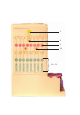
1 Inner glial limiting membrane
2 Layer of nerve fibers
3 Ganglion cell
4 Ganglionic cell layer
5 Inner plexiform layer
6 Inner nuclear layer
7 Outer plexiform layer
8 Amacrine cell
9 Bipolar ganglion cell
10 Horizontal cell
11 Outer nuclear layer
12 Cone cell
13 Rod cell
14 Layer of rods and cones
15 Pigmented layer
16 Choroid
17 Sclera
18 Synapse
19 Axon
20 Nucleus
21 Myoid
22 Ellipsoid
23 Inner segment
24 Cilium
25 Outer segment
26 Pigment cell
27 Outer glial limiting membrane