
User`s guide


Contents ▲ 235 ▼ Index
Evaluating Apoptosis Assays
The apoptosis assay can be used to examine how many apoptotic cells are within a living
cell population. Dead (or necrotic) cells can be excluded from the evaluation.
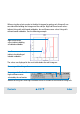
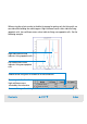
For a detailed description on how to evaluate the results using histograms and regions,
refer to “Using Histograms for Evaluation” on page 199 and “Using Dot Plots for
Evaluation” on page 220.
Living or dead cells
In most cases, you want to know whether cells are dead or alive at a specific time. For
this, you can use calcein-AM as living cell dye, for example. This dye accumulates in
intact cells, whereas it will leak out of damaged cells. Once inside the cells, the colorless
AM ester is cleaved by esterases, resulting in the formation of the highly fluorescent
calcein. The number of events resulting from a calcein-related staining thus gives you the
number of living cells in a sample. For detailed information, refer to the application note
Apoptosis Detection by Annexin V and Active Caspase 3 with the Agilent 2100
Bioanalyzer.
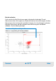
Apoptotic cells
In apoptotic cells, phosphatidylserine is no longer confined to the inner leaflet of the
plasma membrane bilayer. Phosphatidylserine becomes accessible on the outer surface
of the cell membrane and can be bound with high affinity by the protein annexin V, which
can be labeled with biotin or dyes such as Cy5.
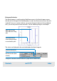
Gating direction
The gating direction is from blue fluorescence (living cells) to red fluorescence (annexin).