
User`s guide

Additional Imaging Modes 7
Agilent 5500 SPM User’s Guide 120
1 nA/V (blue) or 0.1 nA/V (green). See Table 2 on page 115 for more
details.
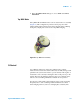
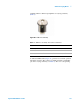
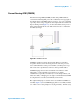
Figure 86 CSAFM nose assembly and scanner
Platinum-coated, conductive tips are required for CSAFM imaging.
Because an electrode must be attached to the sample, a sample plate is
also required.
To image in CSAFM Mode:
1 Begin with the steps you learned in Chapter 4:
a Insert the nose assembly into the scanner.
b Load a probe into the nose assembly.
c Place the scanner in the microscope base and connect its cables.
d Align the laser on the cantilever.
e Insert and align the detector.
2 Prepare the sample and place it on a sample plate. The sample must
be electrically isolated from the sample plate. The particular
mounting arrangement will depend on the sample type and size.
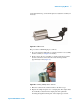
3 Attach an electrode from the sample plate to the sample. A length of
copper wire works well as the electrode. Lift the spring-loaded
electrode clip on the sample plate and insert the electrode under it
(Figure 83 on page 117). Connect the electrode to the sample. Check
the continuity between the working electrode contact and sample to
ensure that a proper connection is achieved.
4 Place the sample plate on the microscope.
5 Plug the 3-pin EC connector of the EC/MAC cable into the 3-pin
socket on the sample plate. Plug the other end of the cable into the
EC/MAC socket on the microscope.