
User`s guide

Additional Imaging Modes 7
Agilent 5500 SPM User’s Guide 130
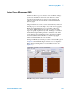
Electrostatic Force Microscopy (EFM)
Electrostatic Force Microscopy (EFM) is a qualitative method for
examining changes in the intrinsic or applied electrostatic field of a
sample surface. EFM is a derivative of AAC Mode, using a conductive
tip. A bias voltage is applied to the sample, allowing local static charge
domains and charge carrier density to be measured. EFM has proven
useful for examining fuel cells, solar cells, and for troubleshooting
semiconductor circuits to locate leaks and shorts.
EFM Mode requires a MAC III controller to provide the drive signals.
Lock-in 1 is used to drive the cantilever. The input to Lock-in 1 is the
amplitude of the cantilever deflection at a specific frequency. Lock-in 2
operates at a different frequency, providing the AC bias, also with the
Deflection channel as its input.
An AC nose assembly and any sample plate with an electrode
connection are required. Conductive EFM tips with a resonance of
approximately 60 kHz are required.
The phase of the Lock-in 2 signal changes in response to changes in the
electric field as the tip passes over the surface. The real component of
the phase (X Component 2) and the total phase can both be mapped. A
standard topography image can be collected at the same time. The two
images can then be displayed side-by-side to highlight correlation
between the electrostatic response and topography.
1 To image in EFM Mode, first follow the steps from Chapter 4:
a Insert the nose assembly into the scanner.
b Insert a probe into the nose assembly.
c Place the scanner in the microscope base.
d Align the laser on the cantilever.
e Insert and align the detector.
2 Prepare the sample and place it on a sample plate. The sample must
be electrically isolated from the sample plate.
3 Attach a conductor (typically a stiff wire) from the working electrode
to the sample. Lift the spring-loaded electrode clip on the sample
plate and insert the conductor under it. Connect the conductor to the
sample. Check the continuity between the working electrode contact
and sample to ensure a good connection.
4 Plug the 3-pin EC connector of the EC/MAC cable into the 3-pin
socket on the sample plate. Plug the other end of the cable into the
EC/MAC socket on the microscope.
5 Choose Mode > EFM.