
User's Manual


Acclarix AX3 Series Diagnostic Ultrasound System User Manual Ultrasound Intensity and Safety
- 133 -
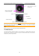
B.3.3 Display of MI/TI
The system provides real-time display of MI/TI values in the upper right part of the screen. The start
point of MI/TI value is 0.0.
The operator should monitor these values during examinations and keep the exposure time and
output level at the minimum amounts needed for effective diagnosis.
The display precision is 0.1.
MI display error:
When measured MI≤0.5, the absolute display error≤0.25;
When measured MI﹥0.5, the relative display error ≤±50%.
TI display error:
When measured TI ≤2.0, the absolute display error ≤1.0;
When measured TI﹥2.0, the relative display error ≤±50%.
B.4 Acoustic Output
B.4.1 Factors that Contribute to Uncertainty in the Output Display
A number of factors should be considered in display accuracy determination methods, such as:
Transducer variability
System variability
Measurement variability and accuracy
The number of operating conditions of which the system is capable and the number tested in
obtaining display accuracy results
Whether display accuracy will be determined by specific combinations of system, mode,
transducer assembly and transmit patterns, or all allowed combinations of them
Accuracy of system software MI and TI calculation algorithms.
Engineering approximations for real-time calculations
B.4.2 Differences between Actual and Displayed MI/TI
Actually, many assumptions adopted in the process of measurement and calculations are relatively
conservative. Over-estimation of actual in situ intensity exposure, for the majority of tissue paths, is
made to the measurement and calculation process. For example, attenuation coefficient of 0.3
dB/cm∙MHz, which is much lower than the actual value for most tissues of the body, is adopted. And
conservative values of tissue characteristics are selected for use in TI models. Therefore, the display
of MI and TI should be used as relative information to assist operator in prudent use of ultrasound
system and implementation of ALARA principle, and the values should not be interpreted as the actual
physical values in tissues or organs examined.
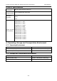
B.4.3 Measurement Uncertainty
The uncertainties in the measurements were predominantly systematic in origin; the random
uncertainties were negligible in comparison. So the intensity and pressure measurement uncertainties
were showed in table 1and table 2. The power, the center frequency and MI measurement
uncertainties were showed in table 3.