
Operating Manual


utilizing DDAs for radiographic inspection versus traditional film and computed radiography
techniques. The reduction in exposure time not only enables productivity through shorter shot
times, instant availability of images for review and analysis; but also improves overall safety to
radiation workers and other employees by decreasing radiation source deployment or on-time
and in some cases allows for a decrease in energy or source strength.
2.1.1 Application Example
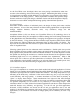
There are many examples where DDAs have shown significant benefits over film or
computed radiography. One of these examples is in the oil and gas downstream inspection
of valves. The same inspection plan was completed utilizing both computed radiography
and a DDA. The image quality results were similar or improved utilizing the DDA, but the
exposure time results were remarkably reduced.
Figure 1: Two application examples showing comparing DDA and CR exposure times
2.2 History of DDA Innovation
The medical industry began developing DDA technology over 25 years ago, spending
millions of dollars in the initial 10-year development cycle. Since initial introduction,
technology investment has continued, focusing on two critical areas: image quality for
visualization of relevant features and dose reduction for improved doctor, healthcare
worker, and patient safety. The focus on optimization of image quality with respect to
dose is one of the key aspects leading to the successful implementation into industrial field
applications, where radiation safety is a critical consideration versus environments where
shielding and radiation protection cabinets can be utilized.
2.3 DDA Design Considerations