
user manual


3
3 - 1
3. OPERATION
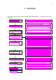
Shown below is the procedural flow of typical S-3400 SEM operation. For details, refer to each
subsection.
●
Start-up Confirmation Items (3.1.1)
●
Startup Operation (3.1.2)
●
Precautions Concerning Specimen Preparation (3.2.1)
●
Specimen Preparation according to Material (3.2.2)
●
Mounting a Specimen on the Type I Stage (Manual Stage)
(3.2.3)
●
Mounting a Specimen on the Type II Stage (5-axis motor-driven stage)
(3.2.4)
●
Conditions under which Accelerating Voltage can be Applied
(3.3.1)
●
Setting the Accelerating Voltage and Filament Current (3.3.2)
●
Setting Parameters for the Electron Optical System (3.4.1)
●
Axial Alignment (3.4.2)
● Selecting a Detector (3.5.1)
● Selecting Magnification (3.5.2)
● Selecting Scanning Speed (3.5.3)
● Image Brightness and Contrast Adjustment (3.5.4)
● Focus and Astigmatism Correction (3.5.5)
●
Operation of the Specimen Stage (Type I - Manual Stage)
(3.5.6)
●
Operation of the Specimen Stage (Type II - 5-Axis Motorized Stage)
(3.5.7)
●
Saving and Recording Images (3.6.1)
●
Preparing Images for Recording (3.6.2)
●
Image Capture (3.6.3)
●
Saving a Scanning Image (Direct Save) (3.6.4)
●
Saving Captured Images (3.6.5)
●
Taking Photographs (Option) (3.6.6)
●
Recording Data Display with Images (3.6.7)
●
Turning High Voltage Off (3.8.1)
●
Setting the Stage at the Specimen Exchange Position (3.8.2)
●
Withdrawing the Specimen (3.8.3)
●
Closing Windows and Shutting off the Display Power (3.8.4)
●
Termination Confirmation Items (3.8.5)
●
Precaution on SEM Data Manager
(3.11.1)
●
Functions (3.11.2)
●
Operation (3.11.3)
3.2 Mounting a Specimen
3.3
A
ppl
y
in
g
the Acceleratin
g
Voltage
3.4 Adjusting the Electron
Optical System
3.5 Operation for Image
Observation
3.6 Saving and Recording
Images
3.8 Shutting Down
3.9 Other Functions
3.10 Image Quality
3.1 Starting the System
3.7 Using SEM Data Manager