
Use Instructions


PN 10232 Eagle V1.2 Instructions for Use Rev 1.0 Page 17 of 101
2.1. HCF – Handheld Fluorescence Camera
The Handheld Fluorescence Camera (HFC) is the primary element of the Eagle V1.2 Imaging System.
The HFC is a portable, handheld and battery-
operated imaging device, equipped with an
AMOLED color display screen, and an optical head
integrated with excitation and emissions optics to
enter and image the surgical cavity.
The optical head contains an ambient light sensor
to detect ambient lighting conditions described in
6.2.2.2.5.1, and a range finder to detect imaging
range as described in section 6.2.2.2.2.
Located in the center above the display screen,
the HFC has a single power button to turn the
device on, and to sleep or wake the display screen
once powered on. Refer to section 6.2.1.1 for full
details.
Located on the left of the display screen, the HFC
has a camera and video button, used to
capture and save images and videos respectively. Refer to section 6.2.1 for full details.
Located on the right of the display screen, the HFC has White Light and Fluorescence buttons, to
enable white light LEDS for White Light Imaging or blue-violet LEDS for Fluorescence imaging, respectively.
For full details refer to section 6.2.2.2.4 for White Light Imaging and section 6.2.2.2.5 for Fluorescence
Imaging.
The HFC lithium ion battery is rechargeable using the Contact Charging Device (CCD) described in Section
2.2.5 and Section 6.3.5.
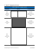
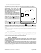
2.1.1. HFC Optical Head
The HFC optical head shown in Figure 7 integrates two camera sensors, one dedicated for White Light
Imaging and one dedicated for Fluorescence Imaging. During White Light Imaging described in section
6.2.2.2.4, two white light LEDs illuminate the field of view. During Fluorescence Imaging described in
section 6.2.2.2.5, four blue-violet light LEDs illuminate the field of view.
2.1.1.1. Emissions Filter
A custom infrared emission filter (95% Transmission of wavelengths 420 – 700 nm and 815 – 900 nm and
blocking all other NIR light >700nm) placed in front of the white light camera sensor ensures only light in
the visible spectrum is captured.
A custom optical emission filter (>95% transmission of wavelengths 500 – 545 nm and 600 – 665 nm and
blocking NIR light >700nm) placed in front of the fluorescence camera ensures only the resulting red
fluorescence emission from PpIX in malignant tissues, and green fluorescence emission from connective
tissues and stroma are captured.
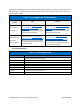
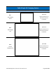
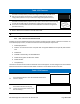
Figure 6 Handheld Fluorescence Camera – HFC
Camera
/ Video
Capture
Buttons
Power Button
Optical Head
WL/FL Buttons
Display Screen