
Use Instructions


PN 10232 Eagle V1.2 Instructions for Use Rev 1.0 Page 74 of 101
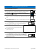
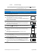
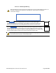
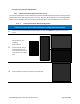
6.2.2.2.5.2. Interpretation of Fluorescence Images
Table 38 provides exemplary white light and fluorescence images of grossly sectioned breast tissue
specimens.
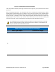
Areas of red PpIX fluorescence are contrasted with areas of healthy tissue autofluorescence which
typically appear predominately green and brown. Areas containing connective tissue typically appear
green fluorescent. Areas composed predominately of adipose tissue typically appear dull pink or brown.
Tissue specimen ink applied to tissue margins typically appears black. Blood vessels and blood also
typically appear black; however, they may also have a dark red hue. Areas of white indicate pixel
saturation and should not be considered when interpreting the images for PpIX fluorescence.
The Handheld Fluorescence Camera utilizes auto-exposure to optimize the exposure settings
based on lighting conditions. Images captured on the HFC are relative, not quantitative, and
may not represent absolute fluorescence levels.
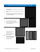
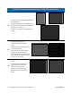
Table 38 Interpretation of Fluorescence Images
White Light Images Fluorescence Images
1.