
Use Instructions

PN 10232 Eagle V1.2 Instructions for Use Rev 1.0 Page 8 of 101
tissue specimens and surgical cavity, beyond what can be visualized based on conventional surgical and
imaging approaches, would provide surgeons with clinically useful real-time information. In patients
undergoing breast conserving surgery, the presence of cancer in the margin of the resected lumpectomy
and/or in the surgical cavity that is undetected by standard of care is a significant clinical challenge for
surgeons and pathologists, and a risk to patients. The Eagle V1.2 Imaging System, when used in
combination a cancer-specific contrast agent, can address these challenges by enabling real-time
fluorescence visualization of otherwise occult cancerous tissue.
Using blue-violet light excitation (405 nm), the Eagle V1.2 Imaging System can visualize protoporphyrin IX
(PpIX) fluorescence emitted by cancer cells in patients who have received aminolevulinic acid (ALA), a
non-fluorescent prodrug contrast agent, prior to surgery [1]. Metabolism of ALA in the body leads to the
selective accumulation of PpIX in cancer cells. Under the blue-violet excitation light emitted by the Eagle
V1.2 Imaging System, the red PpIX fluorescence from cancer cells is detected simultaneously against
background normal tissues (which do not produce significant levels of PpIX) autofluorescence thereby
producing a composite fluorescence image (or video) in which PpIX fluorescent cancer cells and tissues
appear red in color in contrast to healthy surrounding tissues comprised primarily of connective tissue,
which appear predominantly green in color; noting that adipose tissue can appear dull-brown in color in
fluorescence images of breast tissues.
1.4.1. ALA and Protoporphyrin (PpIX) Production
The ALA-induced accumulation of PpIX in cancer cells is what enables the Eagle V1.2 Imaging System to
visualize malignant tissues. The following section describes PpIX synthesis in malignant and healthy tissues
and how exogenous administration of ALA leads to selective accumulation of PpIX in cancer cells thereby
producing fluorescence contrast between cancer cells and surrounding healthy tissues using the Eagle
V1.2 Imaging System.
ALA is a naturally occurring, endogenous substance, which belongs to the group of sensitizers used in
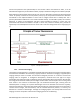
photodynamic diagnosis. It is the first compound in the heme synthesis pathway. Heme biosynthesis
begins within the mitochondrion, where ALA synthase catalyzes the condensation of succinyl-CoA and
glycine to form ALA. Aminolevulinic acid dehydratase (ALAD) condenses 2 molecules of ALA to form the
monopyrrole PBG. PBG deaminase catalyzes the polymerization of 4 molecules of PBG to
hydroxymethylbilane. Hydroxymethylbilane is further metabolized to uroporphyrinogen I and III (by
uroporphyrinogen cosynthase). Uroporphyrinogen decarboxylase sequentially removes a carboxylic
group from the acetic side chains of each of the pyrrole rings to yield coproporphyrinogen.
Coproporphyrinogen oxidase removes a carboxyl group from the propionic groups on 2 of the pyrrole
rings to yield protoporphyrinogen IX. Protoporphyrinogen oxidase forms PpIX by removing 6 hydrogen
atoms from protoporphyrinogen IX. Finally, ferrochelatase mediates the insertion of ferrous iron into the
porphyrin macrocycle, forming heme. Heme biosynthesis is regulated by a negative feedback loop in
which ALA synthase mitochondrial transport is inhibited by heme. Administration of excess exogenous
ALA avoids the negative feedback control, and accumulation of PpIX occurs in target tissue.
ALA HCl for oral solution is usually administered 3 hours (range 2 to 4 hours) before anesthesia prior to
surgery. The investigational drug PD G 506 A is 1.5 g ALA hydrochloride (HCl) granules (chemical name: 5-