
Service manual

103
SpO
2
Accuracy (Mediana module)
The saturation (SpO
2
) accuracy specification was proven by comparisons with the arterial
blood gas measurements. Statistically significant number of samples at SpO
2
levels
ranging from 70% to 99% was collected on male and female volunteers, with different skin
colors.
Pulse rate accuracy specification was proven by laboratory simulator tests, where
oximeter was connected to the Oximetry simulator, set to the precise number of pulses
per minute.
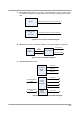
Respiration Processing
The respiration monitoring is designed to use the variation of thoracic impedance. The
chest contains various materials, ranging from bone to air. Each of these materials has
different electrical properties and is located in a different portion of the chest. The
materials of the chest vary in electrical resistivity (the amount of electrical resistance
between opposite faces of a cube of that material), which is an important determinant of
electrical impedance in the body.
Two of the major components of the chest, blood and air, are at opposite ends of the
scale. Furthermore, the volume of each of these materials varies with time over the
cardiac and breathing cycles. The variation of the thoracic impedance is caused by the
difference between air and blood in the thoracic impedance. Blood has relatively low
resistivity, which varies over the cardiac cycle owing to changing blood volumes in the
heart and in the vascular compartment. Air, on the other hand, has high electrical
resistivity and hence impedance, and it undergoes wide volume changes in the lungs
during normal breathing. i.e. the impedance of blood is 150 ohm/cm and the impedance of
air is 5000 ohm/cm.
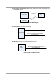
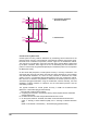
The patient’s respiration is detected by using two of the three leads of the ECG electrodes
(RA and LA, or RA and LL) and cable. The electrical impedance between a pair of
electrodes is determined by dividing the voltage difference between the two electrodes by
the current that passes between them. When the electrodes are placed on the actual
structure, respective structures change.
A low-level excitation signal is applied to these leads, and the variation of the thoracic
impedance caused by breathing is sensed and processed for display and measurement.
This variation is processed to the voltage value for the measurement.
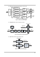
In order to transfer the thoracic impedance by a transformer, it uses a minimum constant
current of the sine wave carrier signal. The transferred thoracic impedance is changed to
the voltage signal by using bridge circuit and differential amplifier. Then, ECG signal is
removed by filter, and carrier frequency is removed by full wave rectifier and filter in order
to extract only thoracic impedance in amplifying at the definite level of signal. This
extracted thoracic impedance signal is used to measure the respiration by digital signal
processing.