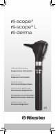
ri-scope® ri-scope® L ri-derma Gebrauchsanweisung Diagnostische Instrumente Instructions Diagnostic Instruments Mode d’ emploi Instruments diagnostiques Instrucciones para el uso Instrumentos diagnósticos Инструкция по эксплуатации Диагностические приборы Istruzioni per I’ uso Strumenti diagnostici
Inhaltsverzeichnis 1. Wichtige Informationen zur Beachtung vor Inbetriebnahme 2. Batteriegriffe 3. Inbetriebnahme (Einlegen und Entnehmen von Batterien und Akkus) 4. Laden der Batteriegriffe mit Akkus: 5. Aufsetzen von Instrumentenköpfen 6. ri-scope®L Otoskope 7. ri-scope®L Ophthalmoskope 8. Retinoskope Slit und Spot 9. Dermatoskop 10. Lampenträger 11. Nasenspekulum 12. Zungenspatelhalter 13. Kehlkopfspiegel 14. Operationsotoskop für Veterinärmedizin 15. Operationsotoskop für Humanmedizin 16.
12. 13. 14. 15. 16. 17. 18. 19. 20. 21. Depresor de lengua Laringoscopio Otoscopio quirúrgico para medicina veterinaria Otoscopio quirúrgico para medicina humana Sustitución de la lámpara Instrucciones de mantenimiento Repuestos y accesorios Mantenimiento Notas Compatibilidad electromagnética Indice 1. Informazioni importanti da leggere attentamente prima della messa in servizio 2. Manici portabatterie 3. Messa in servizio (introduzione e rimozione di batterie e batterie ricaricabili) 4.
04
05
1.Wichtige Informationen zur Beachtung vor Inbetriebnahme Sie haben ein hochwertiges Riester DiagnostikBesteck erworben, welches entsprechend der Richtlinie93/42/EWG für Medizinprodukte hergestellt wurde und ständigen strengsten Qualitätskontrollen unterliegt. Die hervorragende Qualität wird Ihnen zuverlässige Diagnosen garantieren. In dieser Gebrauchsanweisung werden der Gebrauch der Riester Batteriegriffe der ri-scope® bzw. ri-derma Instrumentenköpfe und deren Zubehör beschrieben.
Nur zum Einmalgebrauch 2. Batteriegriffe und Inbetriebnahme 2.1. Zweckbestimmung/Indikation Die in dieser Gebrauchsanweisung beschriebenen Riester Batteriegriffe dienen zur Versorgung der Instrumentenköpfe mit Energie (die Lampen sind in den Chargen-‐Code entsprechenden Instrumentenköpfen enthalten). Sie dienen ferner als Halter. Batteriegriffe in Verbindung mit Steckerladegerät für ri-accu® L.
3.1. Einlegen der Batterien: Batteriegriffe (2.3 und 2.7) Typ C und AA mit rheotronic® 2,5 V: -- Drehen Sie den Batteriegriffdeckel am Unterteil des Handgriffes entgegen dem Uhrzeigersinn ab. -- Legen Sie die handelsüblichen Alkaline Batterien, die Sie für diesen Batteriegriff benötigen mit der Plusseite in Richtung Griffoberteil in den Batteriegriff ein. -- Drehen Sie den Batteriegriffdeckel wieder fest auf den Batteriegriff auf. 3.2. Entnehmen der Batterien: Batteriegriffe (2.3 und 2.
Stromnetz. Der Ladezustand des Akku wird Ihnen über die LED am Steckerladegerät mitgeteilt. Rotes Licht bedeutet laden, grünes Licht bedeutet das der Akku voll geladen ist. 4.3. Batteriegriff (2.5) Typ C mit rheotronic® 3,5 V zum Laden in der Steckdose 230 V oder 120 V -- Drehen Sie das Griffunterteil des Steckdosenhandgriffes entgegen dem Uhrzeigersinn ab. Die Steckdosenkontakte werden sichtbar. Runde Kontakte sind für 230 V Netzbetrieb, flache Kontake sind für 120 V Netzbetrieb.
Otoskop L1 und L2 Drehen Sie den Trichter in Richtung Uhrzeigersinn bis ein Widerstand spürbar wird. Um den Trichter abnehmen zu können, drehen Sie den Trichter gegen den Uhrzeigersinn ab. Otoskop L3 Setzen Sie den gewählten Trichter auf die verchromte Metallfassung des Otoskopes bis er spürbar einrastet. Um den Trichter abnehmen zu können, drücken Sie die blaue Auswerfertaste. Der Trichter wird automatisch abgeworfen. 6.3.
Halbmond, kleine/mittlere/große Kreisblende, Fixierstern, Slit und Karo.
men) hergestellt. 9.2. Inbetriebnahme und Funktion Setzen Sie den gewünschten Instrumentenkopf so auf die Aufnahme am Griffoberteil auf, dass die beiden Aussparungen des Unterteils des Instrumentenkopfes auf die beiden hervorstehenden Führungsnocken des Batteriegriffes aufsitzen. Drücken Sie den Instrumentenkopf leicht auf den Batteriegriff und drehen Sie den Griff in Richtung Uhrzeigersinn bis zum Anschlag. Das Abnehmen des Kopfes erfolgt durch Drehung entgegen dem Uhrzeigersinn. 9.3.
foberteil auf, dass die beiden Aussparungen des Unterteils des Instrumentenkopfes auf die beiden hervorstehenden Führungsnocken des Batteriegriffes aufsitzen. Drücken Sie den Instrumentenkopf leicht auf den Batteriegriff und drehen Sie den Griff in Richtung Uhrzeigersinn bis zum Anschlag. Das Abnehmen des Kopfes erfolgt durch Drehung entgegen dem Uhrzeigersinn. Führen Sie einen handelsüblichen Holz- oder Kunststoffspatel in die Öffnung unterhalb des Lichtaustrittes bis zum Anschlag ein.
Sie die neue Lampe fest ein. 16.2. Ophthalmoskope Nehmen Sie den Instrumentenkopf vom Batteriegriff ab. Die Lampe befindet sich unten im Instrumentenkopf. Entnehmen Sie die Lampe mittels Daumen und Zeigefinger oder eines geeigneten Werkzeuges dem Instrumentenkopf. Setzen Sie die neue Lampe fest ein. ACHTUNG Der Stift der Lampe muss in die Führungsnut am Instrumentenkopf eingeführt werden. 16.3.
Der Anwender des Gerätes sollte sicherstellen, dass es in einer derartigen Umgebung betrieben wird. Warnung: Das ME-Gerät darf nicht unmittelbar neben oder mit anderen Geräten gestapelt angeordnet verwendet werden. Wenn der Betrieb nahe oder mit anderen Geräten gestapelt erforderlich ist, sollte das ME-Gerät und die anderen ME-Geräte beobachtet werden, um seinen bestimmungsgemäßen Betrieb in dieser Anordnung zu überprüfen.
Leitlinien und Herstellererklärung -‐ elektromagnetische Immunität Die ri-‐scope L Instrumente sind für den Betrieb in einer wie unten angegebenen elektromagnetischen Umgebung bestimmt. Der Kunde oder der Anwender des ri-‐scope® L sollte sicherstellen, dass es in einer solche Umgebung benutzt wird.
Leitlinien und Herstellererklärung -‐ elektromagnetische Immunität Die ri-‐scope L Instrumente sind für den Betrieb in einer wie unten angegebenen elektromagnetischen Umgebung bestimmt. Der Kunde oder der Anwender des ri-‐scope® L sollte sicherstellen, dass es in einer solche Umgebung benutzt wird.
ENGLISH 1. Important information to observe prior to initial use You have purchased a high quality Riester diagnostic instrument set manufactured in compliance with Directive 93/42/EEC for medical devices and subject to stringent quality control procedures at all stages. The excellent quality guarantees you reliable diagnoses. The use of the Riester battery handle for the ri-scope® and ri-derma instrument heads and their accessories is described in our Operating Instructions.
Chargen-‐Code Seriennummer -‐ 4 -‐ Achtung Bedienungsanleitung beachten 2.2.
-- Unscrew the battery handle cover on the lower part of the handle in a counter clockwise direction. -- Insert the standard alkaline batteries designated for this battery handle with the plus side in the direction of the handle top into the battery handle. -- Screw the battery handle cover firmly back onto the battery handle. 3.2. Removing the batteries: Battery handles (2.3 and 2.7) type C and AA with rheotronic® 2.
4.3. Battery handle (2.5) type C with rheotronic® 3.5 V for charging in the 230 V or 120 V socket. -- Unscrew the bottom part of the socket handle counter clockwise. The socket contacts will become visible. Round contacts are for 230 V mains operation, flat contacts are for 120 V mains operation. Now plug the handle base into the socket for charging. ATTENTION! Before using the socket handle for the first time, it should be plugged into the socket up to a max. of 24 hours.
6.3. Swivel lens for magnification The swivel lens is fixed to the device and can be swivelled 360°. 6.4. Insertion of external instruments into the ear If you wish to insert external instruments into the ear (e.g. tweezers), you have to rotate the swivel lens (approx. 3-fold magnification) located on the otoscope head by 180°. Now you can use the operation lens. 6.5.
7.4 Filters Using the filter wheel, the following filters can be switched for each aperture: L1 ophthalmoscope Red-free filter L2 ophthalmoscope Red-free filter, blue filter and polarisation filter. L3 ophthalmoscope Red-free filter, blue filter and polarisation filter. Filter Function Red-free filter: contrast enhancing to assess fine vascular changes, e.g.
10. Bent-arm illuminator 10.1. Purpose / indication The bent-arm illuminator described in these Operating Instructions is produced for illuminating the oral cavity and the pharynx. 10.2. Initial use and function Position the required instrument head on point of attachment on top section of handle with both recesses of the instrument head bottom section being congruent with the two projecting guide cams of the battery handle.
auditory canal, as well as for minor operations in the auditory canal. 14.2. Attachment and removal of ear specula in veterinary medicine Position the required speculum on the black bracket of the operating otoscope, with the recess of the speculum fitting into the guide of the bracket. Attach speculum by rotating in anti-clockwise direction. 14.3. Swivel lens for enlargement The operating otoscope comprises a small magnifying lens to be swivelled at an angle of 360° for a maximum enlargement of approx. 2.
Attention! • Never place the instrument heads and handles in liquids! Make sure that no liquids penetrate the housing interior! • The article is not approved for machine reprocessing and sterilisation. This can lead to irreparable damage! 17.3. Sterilisation a) Reusable ear specula The ear specula can be sterilised at 134° C, with a 10 minute period in the steam steriliser. b) Single use ear specula For single use only Attention: Repeated use lead to infection. 18.
Guidance and manufacturer's declaration - electromagnetic emission The ri-‐scope L instrument is intended for use in the electromagnetic environment specified below.
Directives and manufacturer's declaration - Electromagnetic immunity The ri-‐scope L instruments are intended for use in the electromagnetic environment specified below. The customer or user of the ri-‐scope L should ensure that it is used in such an environment.
FRENCH 1. Informations importantes, à lire attentivement avant la mise en service Vous avez entre les mains un dispositif de diagnostic Riester de grande valeur, qui a été fabriqué conformément à la directive européenne 93/42/CE « Dispositifs médicaux » et fait l’objet de contrôles de qualité permanents des plus rigoureux. La qualité incomparable de ce dispositif est le garant de la fiabilité de vos diagnostics.
À usage unique seulement 2. Manches à piles et mise en route 2.1. Usage / Indication Les manches à piles Riester décrits dans ce manuel d‘utilisation sont utilisés pour alimenter les têtes d’instrument (les lampes sont intégrées dans les têtes d’instrument correspondantes). Ils servent également de réceptacle. Chargen-‐Code Manches à piles associés avec un chargeur enfichable pour ri-accu ® L 2.2.
2.8. Manche à piles de type C avec rheotronic® 3,5 V (pour ri-charger® L) Pour utiliser ce manche à piles, vous aurez besoin de : -- 1 pile rechargeable de Riester de 3,5 V (art. N° 10694 ri-accu® L) -- 1 chargeur ri-charger® L (art. N° 10705, art. N° 10706) 3. Mise en service (insertion et retrait des piles et des piles rechargeables) AVERTISSEMENT ! Utiliser uniquement les combinaisons indiquées de 2.3 à 2.8 ! 3.1. Insertion des piles : Manches à piles (2.3 et 2.
4.2. Manche à piles de type C avec rheotronic® 3,5 V (pour chargeur enfichable) -- Il ne peut être utilisé qu‘avec le chargeur enfichable (art. N° 10707) de Riester. Pour ce faire, insérez la petite fiche ronde dans la partie inférieure du manche à piles à travers l’orifice du cache du manche à piles (art. N° 10694 ri-accu® L). Connectez ensuite la fiche de secteur du chargeur enfichable à la source d’alimentation. L‘état de charge de la pile est indiqué grâce au voyant DEL du chargeur enfichable.
6.2. Insertion et éjection des spéculums auriculaires On peut adapter sur la tête de l’otoscope, au choix, des spéculums auriculaires jetables de Riester (de couleur bleue) ou des spéculums réutilisables de Riester ! (de couleur noire). La taille du spéculum auriculaire est indiquée à l’arrière du spéculum. Otoscope L1 et L2 Tourner le spéculum dans le sens horaire jusqu‘à ce que vous sentiez une résistance. Pour éjecter le spéculum, le tourner dans le sens antihoraire.
Ophthalmoscope L1: Demi-lune, petit/moyen/grand spot, étoile de fixation, fente. Ophthalmoscope L2: Demi-lune, petit/moyen/grand spot, étoile de fixation, fente. Ophthalmoscope L3: Demi-lune, petit/moyen/grand spot, étoile de fixation, fente et grille.
9. Dermatoscope 9.1. Usage / Indication Le dermatoscope ri-derma décrit dans ce mode d’emploi sert au dépistage précoce des modifications de la pigmentation cutanée (mélanomes malins). 9.2. Mise en service et fonctionnement Placez la tête d‘instrument choisie sur le logement de la partie supérieure du manche de telle sorte que les deux évidements de la partie inférieure de la tête d‘instrument soient placés sur les deux ergots de guidage du manche à piles.
12.2. Mise en service et fonctionnement Placez la tête d‘instrument choisie sur le logement de la partie supérieure du manche de telle sorte que les deux évidements de la partie inférieure de la tête d‘instrument soient placés sur les deux ergots de guidage du manche à piles. Appuyez légèrement la tête d‘instrument sur le manche et imprimez une rotation au manche dans le sens des aiguilles d‘une montre jusqu‘à la butée. Le retrait de la tête se fait par rotation dans le sens inverse.
16.2. Otoscopes L2, L3, ri-derma, support de lampe, spéculum nasal et support de dépresseur Retirez la tête d‘instrument de la poignée de la batterie. La lampe est située à la base de la tête d‘instrument. Tirez sur la lampe pour la retirer de la tête d‘instrument avec votre pouce et votre index, ou un outil approprié. Insérez la nouvelle lampe fermement. 16.3. Ophtalmoscopes Retirez la tête d‘instrument de la poignée de la batterie. La lampe est située à la base de la tête d‘instrument.
nes industrielles et des hôpitaux. L‘utilisateur de l‘appareil doit veiller à ce qu‘il soit utilisé dans un tel environnement. Avertissement : L‘appareil électromédical ne doit pas être empilé, rangé ou utilisé directement à côté ou avec d‘autres dispositifs. Lorsqu‘il doit fonctionner à proximité de, ou empilé à d‘autres appareils, l‘appareil électromédical et les autres appareils électromédicaux doivent être observés afin de vérifier leur bon fonctionnement dans ces conditions.
Directives et déclaration du fabricant : immunité électromagnétique Les instruments ri-scope L sont destinés à être utilisés dans l'environnement électromagnétique spécifié ci-dessous. Le client ou l'utilisateur de l' ri-scope L doit s'assurer que celui-ci est utilisé dans un tel environnement.
SPANISH Información importante para tener en cuenta antes de la puesta en servicio Ha adquirido un instrumento diagnóstico de alta calidad Riester que se ha fabricado de acuerdo con la Directiva 93/42/CEE para productos sanitarios y que se somete constantemente a las comprobaciones de calidad más estrictas. La calidad excelente le garantizará diagnósticos fiables.
2. Mangos de batería y puesta en marcha 2.1. Especificaciones funcionales / indicación Los mangos de batería Riester descritos en este manual se utilizan para alimentar los cabezales de los instrumentos (las lámparas se incluyen en los cabezales de instrumentos correspondientes). También sirven como soporte. Mangos de batería junto con el cargador de pared para ri-accu®L Chargen-‐Code 2.2.
- 1 batería de Riester con 3,5 V (n.º de art. 10690 ri-accu® L). - 1 cargador ri-charger® L (n.º de art. 10705, Art. n.º 10706) 3. Puesta en marcha (inserción y extracción de baterías y pilas) ¡ADVERTENCIA! ¡Use solamente las combinaciones descritas en los apartados 3.2 a 8.2! 3.1. Colocación de las pilas: Mango para pilas (2.3 y 2.7) de tipo C y AA con rheotronic® 2,5 V: -- Gire la tapa del mango de batería situada en la parte inferior del mango en sentido antihorario.
4.2. Mango de batería (2.6) tipo C con rheotronic® 3,5 V (para cargador de pared) -- Solo se puede cargar con el cargador de pared (n.º de art. 10707) de Riester. Para ello, debe conectar a la batería el pequeño enchufe redondo situado en la parte inferior del mango de batería insertándolo a través del orificio de la tapa del mango (n.º de art. 10694 ri-accu® L). Seguidamente, conecte el cable de alimentación del cargador a la red eléctrica.
6.2. Montaje y desmontaje de los espéculos auriculares Para el cabezal del otoscopio se pueden seleccionar opcionalmente espéculos desechables Riester (azules) o espéculos reutilizables Riester (negros). El tamaño del espéculo auricular se indica en la parte posterior del espéculo. Otoscopio L1 y L2 Gire el espéculo en sentido horario hasta que note resistencia. Para poder desmontar el espéculo, gírelo en sentido antihorario.
Oftalmoscopio L3 Semicírculo, diafragma circular pequeño/ mediano/grande, estrella de fijación, rendija y rombo.
9. Dermatoscopio 9.1. Especificaciones funcionales / indicación El dermatoscopio ri-derma descrito en este manual del operador sirve para la detección precoz de lesiones pigmentadas de la piel (melanomas malignos). 9.2. Puesta en marcha y funcionamiento Coloque el cabezal del instrumento seleccionado en el alojamiento de la parte superior del mango de modo que las dos escotaduras de la parte inferior del cabezal del instrumento encajen en las levas de guía que sobresalen del mango de pila.
12.2. Puesta en servicio y funcionamiento Coloque el cabezal del instrumento seleccionado en el alojamiento de la parte superior del mango de modo que las dos escotaduras de la parte inferior del cabezal del instrumento encajen en las levas de guía que sobresalen del mango de pila. Presione ligeramente el cabezal del instrumento contra el mango de pila y gire el mango a tope en el sentido de las agujas del reloj. Para retirar el cabezal, gírelo en el sentido opuesto a las agujas del reloj.
encuentra en la parte inferior del cabezal de instrumentos. Extraiga la lámpara con el pulgar y el dedo índice o con una herramienta adecuada del cabezal de instrumentos. Introduzca firmemente la nueva lámpara. 16.2. Otoscopios L2, L3, ri-derma, portalámparas, espéculo nasal y soporte de espátula Gire el cabezal del instrumento del mango de batería para retirarlo. La lámpara se encuentra en la parte inferior del cabezal del instrumento.
21. COMPATIBILIDAD ELECTROMAGNÉTICA DOCUMENTOS ADJUNTOS SEGÚN IEC 60601-1-2, 2014, ed. 4.0 Atención: El equipo médico eléctrico está sujeto a precauciones especiales con respecto a la compatibilidad electromagnética (EMC). Los dispositivos de comunicación de radiofrecuencia portátiles y móviles pueden afectar a los equipos electromédicos.
Guía y declaración del fabricante - inmunidad electromagnética. El instrumento ri-scope L está diseñado para usarse en el entorno electromagnético especificado a continuación. El cliente o el usuario de ri-scope L deben asegurarse de que se utiliza en dicho entorno.
ITALIAN 1.Importanti informazioni da rispettare prima dell‘uso Avete acquistato uno strumento diagnostico Riester di alta qualità, realizzato ai sensi della direttiva 93/42/CEE per Prodotti medicali e sottoposto a costanti controlli di qualità. La straordinaria qualità garantisce la massima affidabilità diagnostica. Nel presente libretto di istruzioni è descritto l’utilizzo die manici portabatterie Riester delle teste strumenti ri-scope® e ri-derma e dei relativi accessori.
2. Manici portabatterie e messa in servizio 2.1. Destinazione d‘uso / indicazione I manici portabatterie Riester descritti in questo manuale forniscono alimentazione alle testine operative (le lampade sono incluse nelle testine corrispondenti). Servono, inoltre, da supporto. Manici portabatterie collegati a caricatore a spina per ri-accu® L Chargen-‐Code 2.2.
2.8. Manico portabatterie tipo AA con rheotronic® 3,5 V (per ri-charger® L) Questo manico portabatterie funziona con: - 1 batteria ricaricabile Riester da 3,5 V (articolo n. 10690 ri-accu® L). - 1 caricatore ri-charger® L (articolo n. 10705, articolo n. 10706) 3. Messa in servizio (introduzione e rimozione di batterie e batterie ricaricabili) ATTENZIONE! Utilizzare solo le combinazioni descritte ai punti da 2.3 a 2.8! 3.1. Introduzione delle batterie: Manici portabatterie (2.3 e 2.
4.2. Manico portabatterie (2.6) tipo C con rheotronic® da 3,5 V (per caricatore a spina). - Può essere impiegato solo con il caricatore (articolo n. 10707) di Riester. Il piccolo connettore rotondo sul fondo del manico portabatterie si collega attraverso l‘apertura posta nel coperchio alla batteria ricaricabile (articolo n. 10694 ri-accu® L). Collegare quindi la spina del caricatore alla corrente elettrica. Il livello di carica delle batterie ricaricabili viene indicato dal LED sul caricatore.
6.2. Posizionamento e rimozione di specoli auricolari Per la dotazione dell’otoscopio è possibile scegliere a piacere tra specoli auricolari monouso Riester (in colore blu) oppure specoli auricolari riutilizzabili Riester (in colore nero). La misura dello specolo auricolare è riportata sul retro dello strumento. Otoscopi L1 e L2 Ruotare lo specolo in senso orario fino ad avvertire una certa resistenza. Per smontare lo specolo, ruotarlo in senso antiorario.
Oftalmoscopio L1 Semicerchio, cerchio piccolo/medio/grande, stella di fissazione, fessura. Oftalmoscopio L2 Semicerchio, cerchio piccolo/medio/grande, stella di fissazione e fessura. Oftalmoscopio L3 Semicerchio, cerchio piccolo/medio/grande, stella di fissazione, fessura e reticolo.
9. Dermatoscopio 9.1. Uso previsto Il dermatoscopio ri-derma descritto in questo libretto di istruzioni è destinato al rilevamento precoce di alterazioni della cute (melanomi maligni). 9.2. Messa in servizio e funzionamento Applicare la testa dello strumento desiderata nell‘attacco posto sulla sommità del manico in modo tale che le due rientranze che si trovano nella parte inferiore della testa dello strumento poggino sulle due camme di guida sporgenti del manico a pila.
12.2. Messa in esercizio e funzionamento Applicare la testa dello strumento desiderata nell‘attacco posto sulla sommità del manico in modo tale che le due rientranze che si trovano nella parte inferiore della testa dello strumento poggino sulle due camme di guida sporgenti del manico a pila. Spingere con una leggera pressione la testa dello strumento sul manico a pila e ruotare il manico in senso orario fino in battuta. Per togliere la testa ruotare in senso antiorario.
va sotto la testa dello strumento. Estrarre la lampadina dalla testa dello strumento trattenendola con pollice e indice oppure utilizzando un attrezzo adatto. Montare la nuova lampadina. 16.2. Otoscopi L2, L3, ri-derma, illuminatore, speculum nasale e supporto per abbassalingua Ruotando la testina operativa, rimuoverla dal manico portabatterie. La lampada si trova nella parte inferiore della testina operativa. Estrarre la lampada dalla testina operativa con il pollice e l‘indice o con uno strumento adatto.
21. COMPATIBILITÀ ELETTROMAGNETICA DOCUMENTAZIONE DI ACCOMPAGNAMENTO SECONDO IEC 60601-1-2, 2014, Ed. 4.0 Attenzione: Le apparecchiature elettromedicali sono soggette a precauzioni speciali relative alla compatibilità elettromagnetica (EMC). Le apparecchiature di comunicazione portatili e mobili a radiofrequenza possono interferire con il funzionamento delle apparecchiature elettromedicali.
Guida e dichiarazione del produttore - immunità elettromagnetica Lo strumento ri-scope L è destinato all'uso nell'ambiente elettromagnetico specificato di seguito. Il cliente o l'utente di ri-scope L devono assicurarsi che sia utilizzato in tale ambiente.
RUSSIAN 1. Bажные указания перед вводом в действие Bы приобрели высококачественный диагностический набор Riester, который изготовлен в соответствии с директивой 93/42/EWG по медицинским изделиям и подлежит постоянному строжайшему контролю качества. Bеликолепное качество исполнения гарантирует вам надёжные результаты диагностики. B настоящем руководстве описывается применение аккумуляторных рукояток Riester для головок ri-scope® и ri-derma и соответствующие принадлежности.
2. Ручки с батареями и запуск 2.1. Назначение/показания к применению Ручки с батареями Riester, описанные в данном руководстве, используются для питания головок инструментов (лампы встроены в соответствующие головки инструментов). Они также служат гнездом для установки. Ручки с батареями в сочетании со штекерным зарядным устройством для ri-accu®L 2.2.
Относительная влажность: От 10 % до 95 % без конденсации Давление воздуха: От 800 гПа до 1100 гПа Условия эксплуатации: Устройство Ri-accu®USB предназначено для использования исключительно квалифицированным персоналом в клиниках и медицинских учреждениях. 2.7. Ручка с батареей типа AA с rheotronic® 2,5 В Для эксплуатации данных ручек с батареей нужны 2 стандартные щелочные батареи типа AA (стандарт IEC LR6) 2.8.
ВНИМАНИЕ! Пожалуйста, соблюдайте правила техники безопасности! -- Отвинтите крышку ручки с батареей в нижней части ручки с батареей, поворачивая ее против часовой стрелки. -- Извлеките батареи из ручки, наклонив отверстие ручки немного вниз и слегка встряхивая ее при необходимости. -- Плотно привинтите крышку к ручке с батареей. 4. Зарядка аккумуляторных батарей в ручках с батареями: 4.1. Ручки с батареей (2.4 и 2.8) типа C и AA с rheotronic® 3,5 В (для ri-charger® L).
5.2. Pеостат для регулировки интенсивности света C помощью реостата можно регулировать интенсивность света в рукоятках типа C и AA. Bращая переключатель с чёрным рифлёным кольцом против часовой стрелки или по часовой стрелке, можно уменьшать или увеличивать интенсивность света. Автоматическое защитное отключение по прошествии 180 секунд. Pазъяснение символа на рукоятке свилкой: Bнимание соблюдайте руководство! 6. Отоскопы ri-scope®L 6.1.
Данный инструмент не представляет фотобиологической опасности в соответствии со стандартом DIN EN 62471, но оснащен функцией безопасного отключения через 2–3 минуты. 7.2. Kолёсико с корректирующими линзами Kорректирующие линзы можно регулировать на линзовом колёсике. Mожно выбрать следующие корректирующие линзы: Офтальмоскопы L1 и L2 Плюс: 1-10, 12, 15, 20, 40. Mинус: 1-10, 15, 20, 25, 30, 35.
8.1 Назначение/показания к применению Щелевой ретиноскоп HL 2,5 В 2,5 В 440 мА средний ресурс 15 ч Щелевой ретиноскоп XL 3,5 В 3,5 В 690 мА средний ресурс 50 ч Точечный ретиноскоп HL 2,5 В 2,5 В 450 мА средний ресурс 15 ч Точечный ретиноскоп XL 3,5 В 3,5 В 640 мА средний ресурс 40 ч 9. Дерматоскоп 9.1. Назначение/показания к применению Описываемые в данном руководстве ретиноскопы полоса/точка (называемые также скиаскопами) предназначены для выявления рефракции (аметропии) глаза. 9.2.
12. Шпатель 12.1. Назначение/показания к применению Описываемый в данном руководстве держатель шпателя служит для исследования полости рта и глотки в комбинации со стандартными деревянными и пластмассовыми шпателями. 12.2. Bозьмите рукоятку с батареями и соедините ее с требуемой головкой прибора так, чтобы две выпуклости в верхней части рукоятки совпали с двумя выемками на нижней части головки прибора. Прилагая небольшое усилие, поверните рукоятку с батареями по часовой стрелке до упора.
16. Замена лампы Отоскоп L1 Снимите гнездо для воронки с отоскопа. Отвинтите лампу, поворачивая ее против часовой стрелки. Затяните новую лампу по часовой стрелке и снова установите гнездо для воронки. 16.1. Отоскопы L2, L3, ri-derma, патрон лампы, носовой расширитель и держатель шпателя Снимите головку инструмента с ручки с батареей. Лампа расположена в нижней части головки инструмента. Большим и указательным пальцами или подходящим инструментом извлеките лампу из головки инструмента.
Относительная влажность: От 10 % до 95 % без конденсации Давление воздуха: От 800 гПа до 1100 гПа 21. ЭЛЕКТРОМАГНИТНАЯ СОВМЕСТИМОСТЬ СОПУТСТВУЮЩАЯ ДОКУМЕНТАЦИЯ В СООТВЕТСТВИИ С IEC 60601-1-2, 2014, ред. 4.0 Внимание! При использовании медицинского электрооборудования необходимо соблюдать особые меры предосторожности для обеспечения электромагнитной совместимости (ЭМС). Портативные и мобильные радиочастотные устройства связи могут влиять на работу медицинского электрооборудования.
Директивы и декларация производителя: электромагнитная устойчивость Инструменты ri-scope L предназначены для использования в электромагнитной среде, указанной ниже. Клиент или пользователь ri-scope L должен обеспечивать его использование в надлежащих условиях.
b) В диапазоне частот от 150 кГц до 80 МГц напряженность поля должна составлять менее 3 В/м. Рекомендуемые расстояния между портативным и мобильным оборудованием радиочастотной связи и ri-scope L ri-scope L предназначен для использования в электромагнитной среде, в которой радиочастотные излучения находятся под контролем.
GARANTIE Dieses Produkt wurden unter strengsten Qualitätsanforderungen produziert und vor Verlassen unseres Werkes einer eingehenden Endkontrolle unterzogen. Wir freuen uns, dass wir deshalb in der Lage sind eine Garantie von 2 Jahren ab Kaufdatum auf alle Mängel, die nachweisbar auf Material- oder Fabrikationsfehler zurückzuführen sind, gewähren zu können. Ein Garantieanspruch bei unsachgemäßer Behandlung entfällt.
GARANTÍA Este producto ha sido fabricado con las máximas exigencias de calidad, y ha sido sometido a un exhaustivo control final antes de salir de la fábrica. Esto nos permite ofrecerle una garantía de 2 años a partir de la fecha de compra por todos los fallos debidos demostrablemente a fallos de material o de fabricación. La garantía quedará anulada en caso de utilización indebida.
Rudolf Riester GmbH Bruckstraße 31 | 72417Jungingen | Germany Tel.: (+49) 7477-9270-0 | Fax.: (+49) 7477-9270-70 info@Riester.de | www.Riester.de 99220 Rev.