
Operating instructions


CoronaryCTA
195
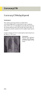
• We recommend using ECG-gated spiral protocols for
optimized image quality of the coronary arteries and
to provide high-quality 3D image data as an input for
3D postprocessing such as MPR, MIP, VRT or Fly
Through. Although ECG-gated spiral scanning is less
sensitive to variable heart rates than ECG-triggered
sequential scanning, the examination of patients
with complex arrhythmia that results in unpredict-
able variations of the RR-intervals (e.g. complex ven-
tricular arrhythmia or multiple extra beats) can result
in limited image quality and should be performed in
exceptional cases only.
• Acquisition with a minimum collimated slice width
ensures best possible image quality due to the opti-
mized intrinsic resolution of the scan data. Once
high quality scan data has been acquired, the recon-
structed slice width has to be optimized with respect
to image noise and best possible quality in MPR, MIP
and VRT reconstructions.